
Brain Tutor 3D
وصف لـBrain Tutor 3D
Explore the brain from the palm of your hand! Learn about the structure and function of the human brain by interacting with high-resolution rotatable 3D models in real-time like you've never experienced it before!
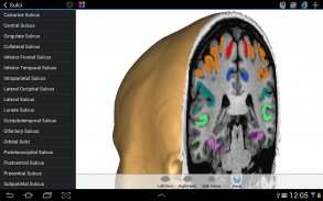
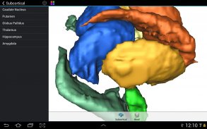
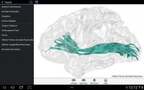
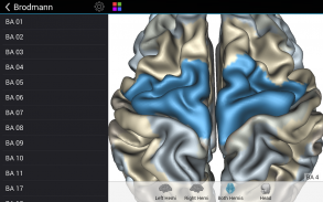
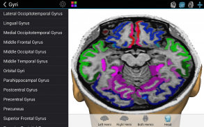
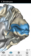
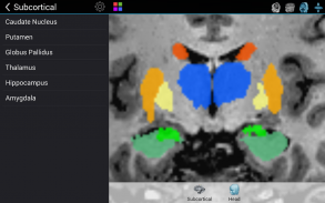
Brain Tutor uses rendered head and brain models as well as fiber tracts that were created from magnetic resonance imaging (MRI) scans of a study volunteer. The MRI data allows to look "inside" the brain using real-time slicing at millimeter resolution. For students, cognitive neuroscientists, medical professionals and everyone interested in the brain, the program provides information about the anatomy and function of the human brain with various atlases describing and visualizing lobes, gyri, sulci, Brodmann areas, subcortical structures, selected specialized functional areas and major fiber tracts.
With Brain Tutor you can:
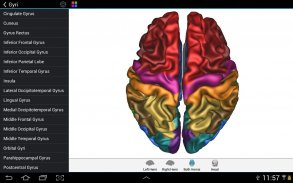
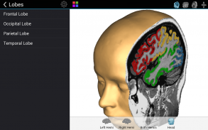
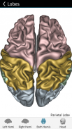
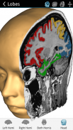
* Explore high-resolution 3D models of the head and brain in real-time.
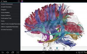
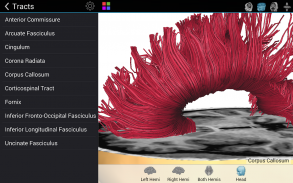
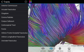
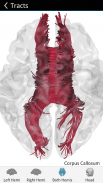
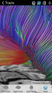
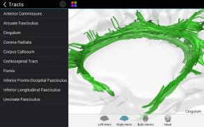
* Visualize major white matter fiber tracts.
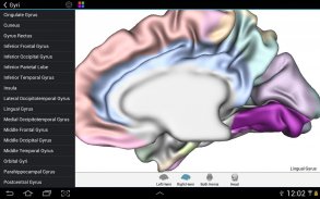
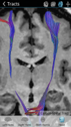
* Slice the brain along three axes (sagittal, axial and coronal).
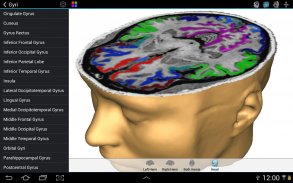
* View MRI brain slices at millimeter resolution.
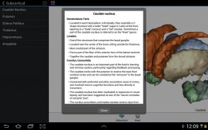
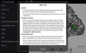
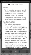
* Learn from text information about the functions of selected lobes, gyri, sulci, subcortical structures, Brodmann areas, functional areas and fiber tracts.
* Learn where brain structures are located both within 3D brain models as well as in MRI slices.
To get started:
* Tap on a 3D brain model to reveal a brain area at that location.
* Switch to another atlas and a specific brain area or fiber tract using the navigation tables.
* Select a 3D model (left/right/both brain hemispheres, head) from the tab bar buttons at the bottom.
* Pan with one finger to rotate a brain model.
* Pan with two fingers to move a brain model.
* Use pinch gesture to zoom a brain model.
* Select the head model to switch to head slicing mode.
* Toggle between navigation and slicing mode by tapping the slicing icon at the top right.
* In head slicing mode, pan with one finger to move the slicing plane through the head.
* Tap on a slicing direction icon in the top bar to switch between three orthogonal slice planes.
* Tap on the displayed name of a selected brain structure to read text information in a popup dialog.
This app has been designed and programmed by Prof. Rainer Goebel, a leading expert in anatomical and functional brain imaging and award-winning developer of scientific software. For more information about his work, see http://www.brainvoyager.com/RainerGoebel.html.
استكشاف الدماغ من كف يدك! التعرف على بنية ووظيفة الدماغ البشري من خلال التفاعل مع عالية الدقة 3D نماذج للتدوير في الوقت الحقيقي وكأنك لم اشهد ابدا من قبل!
يستخدم الدماغ مدرس المقدمة الرأس والدماغ نماذج وكذلك مساحات الألياف التي تم إنشاؤها من التصوير بالرنين المغناطيسي (MRI) بمسح من المتطوعين الدراسة. يسمح للبيانات التصوير بالرنين المغناطيسي للنظر "داخل" الدماغ في الوقت الحقيقي باستخدام تشريح في القرار ملليمتر. للطلاب وعلماء الأعصاب الإدراكي، والمهن الطبية وجميع المهتمين في الدماغ، ويوفر البرنامج معلومات حول تشريح وظيفة من وظائف الدماغ البشري مع مختلف الأطالس التي تصف وتصور الفصوص، التلافيف، تلم، باحات برودمان، وهياكل تحت القشرية، مجالات وظيفية متخصصة مختارة ومساحات الألياف الرئيسي.
مع الدماغ مدرس يمكنك:
* استكشاف نماذج 3D عالية الدقة من الرأس والدماغ في الوقت الحقيقي.
* تصور مساحات الألياف المادة البيضاء الكبرى.
* شريحة الدماغ على ثلاثة محاور (السهمي، محوري والاكليلية).
* عرض التصوير بالرنين المغناطيسي الدماغ شرائح في القرار ملليمتر.
* تعلم من المعلومات حول النص وظائف فصوص المختارة، التلافيف، تلم، وهياكل تحت القشرية، باحات برودمان، مجالات وظيفية ومساحات الألياف.
* تعلم حيث توجد هياكل الدماغ سواء داخل الدماغ نماذج 3D وكذلك في شرائح التصوير بالرنين المغناطيسي.
لتبدأ:
* اضغط على نموذج الدماغ 3D لتكشف عن وجود منطقة في الدماغ في ذلك الموقع.
* التبديل إلى الأطلس آخر ومنطقة محددة في الدماغ أو الألياف المسالك باستخدام الجداول الملاحة.
* حدد نموذج 3D (يسار / يمين / فصي المخ، الرأس) من أزرار شريط التبويب في الجزء السفلي.
* لقطة استعراضية مع إصبع واحد لتدوير نموذج الدماغ.
* لقطة استعراضية مع اثنين من أصابع للتحرك نموذج الدماغ.
* استخدم لفتة قرصة للتكبير نموذج الدماغ.
* حدد نموذج الرأس للتبديل إلى وضع الرأس تشريح.
* تبديل بين الملاحة ووضع تشريح بالنقر على أيقونة التقطيع في اعلى اليمين.
* في وضع تشريح الرأس وعموم مع إصبع واحد لتحريك الطائرة من خلال تشريح الرأس.
* اضغط على أيقونة اتجاه التقطيع في الشريط العلوي للتبديل بين ثلاث طائرات شريحة المتعامدة.
* اضغط على اسم عرض من بنية الدماغ اختيارها لقراءة المعلومات النص في الحوار المنبثقة.
وقد تم تصميم هذا التطبيق ومبرمجة من قبل البروفيسور راينر غوبيل، وهو خبير بارز في التشريحية والوظيفية تصوير الدماغ ومطور البرمجيات العلمية الحائز على جائزة. لمزيد من المعلومات عن عمله، انظر http://www.brainvoyager.com/RainerGoebel.html.
Brain Tutor 3D - إصدار 2.0
(27-02-2023)Brain Tutor 3D - معلومات APK
نُسخة APK: 2.0الحزمة: com.brainvoyager.android.BrainTutorآخر إصدار من Brain Tutor 3D
نُسخ أخرى


 4.34
4.34


 4.83
4.83


تطبيقات من الفئة نفسها